1) Confirm the matrix fits passively over the maxillary centrals
2) While holding the matrix in place, have the patient
"tap" up and
down a few times, to confirm that the Discluding Element contacts
the lower incisor's midline. It may be necessary to move the
matrix
laterally to accomplish this.
3) Reline the matrix with Parkell's SNAP clear, colorless methacrylate
allowing to set up for *2 minutes* only, then remove with a quick motion.
4) Align and orient the matrix so that the lower incisors contact the DE
as close to perpendicular as possible. The bulk of acrylic at labial
side will be reduced and polished to a tapering flush edge. (As
in #12 below)
5, 6) Some occlusal schemes (usually class III) and practitioner's and patient's
preference dictate that the NTI-css matrix be adapted to the lower incisors.
` The same protocol applies.![]()
![]()
*If a patient's habitual movement allows for the lower canine to occlude on the DE
(above left), not only will symptoms continue, but may intensify. Adapting the
NTI device to the mandibular incisors avoids a canine contact (above right). Assuming the
occlusal scheme allows for it, some practitioners choose this as their primary method.
7) Upon removal of the NTI-css matrix, the internal features
resemble
an impression of the teeth.
8) Depending upon the degree of undercuts, considerable
internal relief may be
necessary in order to snap the matrix back onto the teeth.
This step in the protocol makes it obvious that casual dislodgment
of
the device while being worn by the patient is highly unlikely,
and is
an absolute function of the practitioner's care.
![]()
9) Occasionally, one lower central incisor will be taller than the other.
10) If not addressed, the mandible will shift excursively to maintain "equilibration",
that is, equal stimulation of both the right and left mandibular nerve.
11) While the DE can be modified to accommodate in most circumstances,
equilibrating the incisal edges of the incisors is most desirable.

12) The degree of vertical opening that the DE creates may be more than necessary.
In fact, too much vertical opening is contra-indicated. Only a millimeter or two of
disclusion space between the cuspid cusp tips is necessary.
![]()
14) If the DE does create too much vertical opening, it is reduce to allow for
appropriate freeway space between the cuspid cusp tips in excursive movement (15).

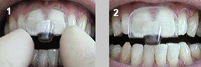
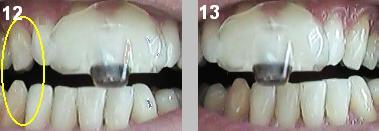
16) In retrusive position, the lower incisors should not be within
2 mm of the distal edge of the DE. If so, gradual resolution of
the patient's myofascial condition may allow for an improved
range of motion, thereby allowing the patient to "bite behind
the bump". (methylmethacrylate is easily adapted on the DE
to enhance it).
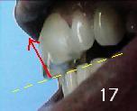
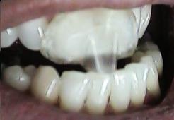
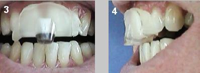
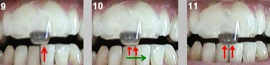
17) Although the angle of the DE is close enough to perpendicular to the
long axis of the lower incisors (where intense clenching does no trauma),
the slope of the DE in #17 above allows for resistance to protrusive/superior
force (red arrow) when the mandible protrudes. The patient's temporal
symtoms may decrease, but facial (lateral ptyergoid) and cervical
(trapizus) symptoms may persist or intensify. (Remedy: Level the DE
parallel the maxially occlusal plane)
![]()
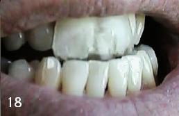
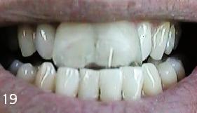
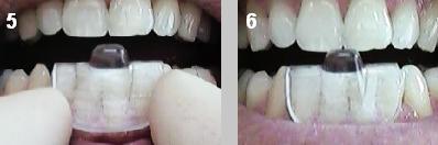
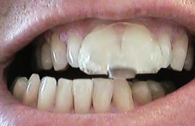
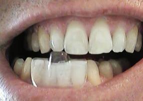
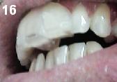
18 and 19: "Getting in Front of the Bump") If the patient can protrude
and place the lower incisors anterior to the labial edge of the DE, it should
lengthened (below) so that in maximum protrusion, the incisors are always contacting
the DE (close to) their long axis. (To confirm whether or not the patient is
actually "getting in front of the bump", ask them to do it while in the office.
If it is uncomfortable, or painful, chances are that they are.)
Facial and TMJ pain typically result if left un-modified.